
Cardiac CT, stress test, EKG…you may have heard these terms before, but do you know what they mean?
These are all types of imaging tests that your provider may order to get pictures of what’s happening in your heart. There are many reasons why you might need a heart imaging test, from experiencing chest pain or shortness of breath to determining whether you’ve had a heart attack to monitoring treatments. Results of imaging tests can help your provider:
- Diagnose things like coronary artery disease, heart failure, or congenital heart defects (problems with how the heart forms before birth)
- Look for buildups of plaque or calcium that may block blood flow
- Discover problems with how your heart or blood valves function
- Visualize how blood flows through your heart and body
- Find signs of heart damage or heart attack
- Prepare for an upcoming heart procedure
The specific test you need depends on your symptoms, the results of previous tests, and the type of test that’s safest for you.
Some people only need one type of test, while others need multiple types. Most of the tests are quick, easy, and painless and don’t require an overnight stay in the hospital.
Also read: Radiology – Types of Imaging Explained
Here is a rundown of some of the most common heart imaging tests.
Electrocardiogram
Electrocardiograms (EKGs, or ECGs) record electrical activity in your heart with ultrasound waves to check your heartbeat. A heartbeat that is too fast or slow can help your provider figure out if there’s an issue. EKGs are often the first test you will need if your provider suspects a heart problem.
This test involves getting sensors (electrodes) placed on your arms, legs, and chest. It may take about 10 minutes to get set up, but the test only takes a few seconds.
Heart CT/Heart Scan
A cardiac computed tomography (CT) scan is an imaging test that uses X‑rays to create detailed pictures of your heart and blood vessels. A computer program then combines the images to make a 3D model of your heart.
During a CT scan, you will lay on a table that slides into the center of a donut-shaped scanner and the x‑ray beams will rotate around you. You will not be enclosed in a machine.
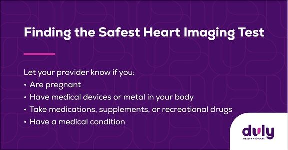
Some heart CTs require contrast, which is a dye that makes your heart and blood vessels appear even clearer in the images. The contrast is given through an IV, so you might feel a tiny pinch when the needle is inserted. You may also feel a warm sensation throughout your body or have a metallic taste in your mouth for a few seconds while the contrast circulates.
The test itself only takes about 10 minutes however you should plan on at least 30 to 60 minutes to include time for preparation.
Cardiac CT Screening
Taking care of your heart health has never been more accessible. Duly Health and Care now offers cardiac CT scans for just $75 at our advanced imaging centers in Lisle and New Lenox. This affordable screening option provides detailed visualization of your heart and blood vessels, helping detect potential issues early when they’re most treatable.
Our state-of-the-art imaging technology and experienced radiology team ensure you receive accurate, high-quality results in a comfortable environment. Whether you’re monitoring an existing condition or being proactive about your cardiovascular health, this cost-effective screening makes it easier to prioritize your heart.
Cardiac MRI
Magnetic resonance imaging (MRI) tests are similar to CT scans, since they also take multiple images of your heart and combine them for a more detailed picture. However, MRIs don’t use X‑rays, so there’s no radiation involved. MRIs take a bit longer than CTs and use a different type of contrast.
When getting an MRI, you will lie down on a table that slides into the MRI machine. As the machine scans your body, it’s important to remain still so that the images are clear. The test can take anywhere from 30 to 90 minutes. You will likely hear loud noises as parts of the machine move around you to take pictures, but you may be able to wear headphones to block them out. Like a CT scan, your provider may need some images with contrast.
Laying still in an enclosed space might make you nervous, especially if you’re claustrophobic (scared of enclosed spaces). But there are microphones and speakers in the machine, so you will be able to communicate with the testing staff if needed. They will also be watching you at all times. Let your provider know ahead of time if you’re feeling anxious – they might be able to give you medication to help you relax.
Also read: Unraveling Cardiology: Breaking Down Subspecialties Within Cardiology
Cardiac Catheterization and Coronary Angiography
Cardiac catheterization is a test where your provider inserts a small tube called a catheter inside your artery and guides it into your heart. There are several types of tests that use catheterization. One of the common ones is a coronary angiography.
During a coronary angiography, your provider will inject dye into the catheter and use X‑rays to see how it moves through your artery. Usually, the test lasts between 30 minutes to an hour.
You will be awake for the procedure and able to follow the provider’s instructions, but there’s no need to worry about pain from the catheter – your provider will give you medicine to prevent pain and to help you relax.
Nuclear Cardiac Stress Testing
Despite what the name suggests, stress testing doesn’t measure how stressed you are – it measures the amount of stress on your heart during exercise or other physical activities. Stress tests can show your provider how well blood pumps through your heart and if your heart receives enough blood from your body. They can also help your provider determine the right amount and type of exercise for you.
Getting a stress test involves a little more effort on your part than some of the other tests. While hooked up to equipment that monitors your heart function, you will walk on a treadmill or ride a stationary bike at increasing speed or resistance levels. You may be asked to breathe into a tube during the test. Afterward, you will have your blood pressure and heart rate checked.
Not all stress tests are imaging tests. When your provider wants to get images of your heart while you take a stress test, they can use nuclear cardiac imaging. This is an advanced technique that can show a level of detail that you can’t get from other imaging tests. During a nuclear test, your provider will inject a small, safe amount of a radioactive substance called a tracer into your bloodstream. The tracer emits gamma rays, which a computer uses to create images that evaluate blood flow, heart muscle function, and coronary arteries.
Also read: Innovation in Cardiac Imaging
The nuclear technique isn’t just for stress tests – it may also be used as part of a CT scan, MRI, or PET scan.
Fun Fact: Duly Health and Care’s Nuclear Program was the first one in Illinois to be fully accredited by the Intersocietal Commission for the Accreditation of Nuclear Medicine Laboratories!
Take Action for Your Heart Health Today
Don’t wait for symptoms to worsen. Whichever test your provider orders, make sure to follow through. Getting tested can help them find problems, get you started on treatment early, and possibly prevent heart issues in the future.
At Duly Health and Care, many of our imaging tests are available at each of our Radiology locations or take place at our same-day Cardiac Evaluation Center. The CEC center offers many same-day testing options, such as CT scans, exercise and stress testing, and EKGs, when you are experiencing non-life-threatening heart symptoms. Your care team, including a board-certified cardiologist, will carefully review test results and provide a personalized care plan all in one convenient visit.
The first step toward getting an imaging test and learning more about your heart health is talking to your cardiologist. Find a Duly Health and Care cardiologist and schedule an appointment online — your heart will thank you!
Health Topics:
-

I believe health care begins with prevention. Life style and risk factor modification can greatly reduce a patient's need for medical and invasive treatments. When these therapies are needed, however, I encourage my patients to take an active role in their health care and make informed decisions that we both believe to be in their best interest.


