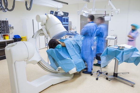
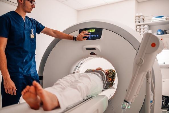
Fluoroscopy, or fluoro, is made up of “live” X‑ray images that when they are put together look like a movie. A fluoroscope allows medical staff to see bones and also helps physicians to identify soft tissue pathology.
Fluoroscopy helps reduce the invasiveness of a surgery. Prior to fluoroscopy, physicians had to surgically open a patient to see the form and function of a certain body part. A large incision through skin and several layers of soft tissue or bone would need to be made to view the anatomy in its entirety. With the help of a fluoroscope, incisions can be smaller, thus greatly shortening recovery time.
Today, many diagnostic and therapeutic exams and surgeries benefit from fluoroscopy. Below are 4 common uses of fluoroscopy:
- A radiologist can use barium to check the functions of the stomach, the small and large intestines, colon and rectum. Since X‑rays often shoot completely through these soft tissues, barium adds density to these anatomies so that they can be monitored.
- During a swallow study, a speech language pathologist can use fluoroscopy to see if food is going to the right place when swallowed. They can also check to see if parts of the patients mouth and throat are working properly.
- In cardiac procedures, dye can be injected into the coronary arteries to show blood flow or to investigate potential blockages. Catheters may be placed more easily due to fluoroscopic guidance.
- Several spine and joint injections can accurately be made using fluoroscopy after dye is injected. These injections can be both diagnostic, to see if there is a greater underlying pathology, or therapeutic, sometimes providing full relief for an extended period of time.
These are just a few of the most common uses and benefits of fluoroscopy.
Health Topics:

